Test for Eye Disease in Hawaii
Managing eye diseases early and correctly is the key to effective treatment. At Retina Consultants of Hawaii, we perform five diagnostic tests — fluorescein angiography, ICG angiography, optical coherence tomography, fundus photography, and ultrasound.
Fluorescein angiography
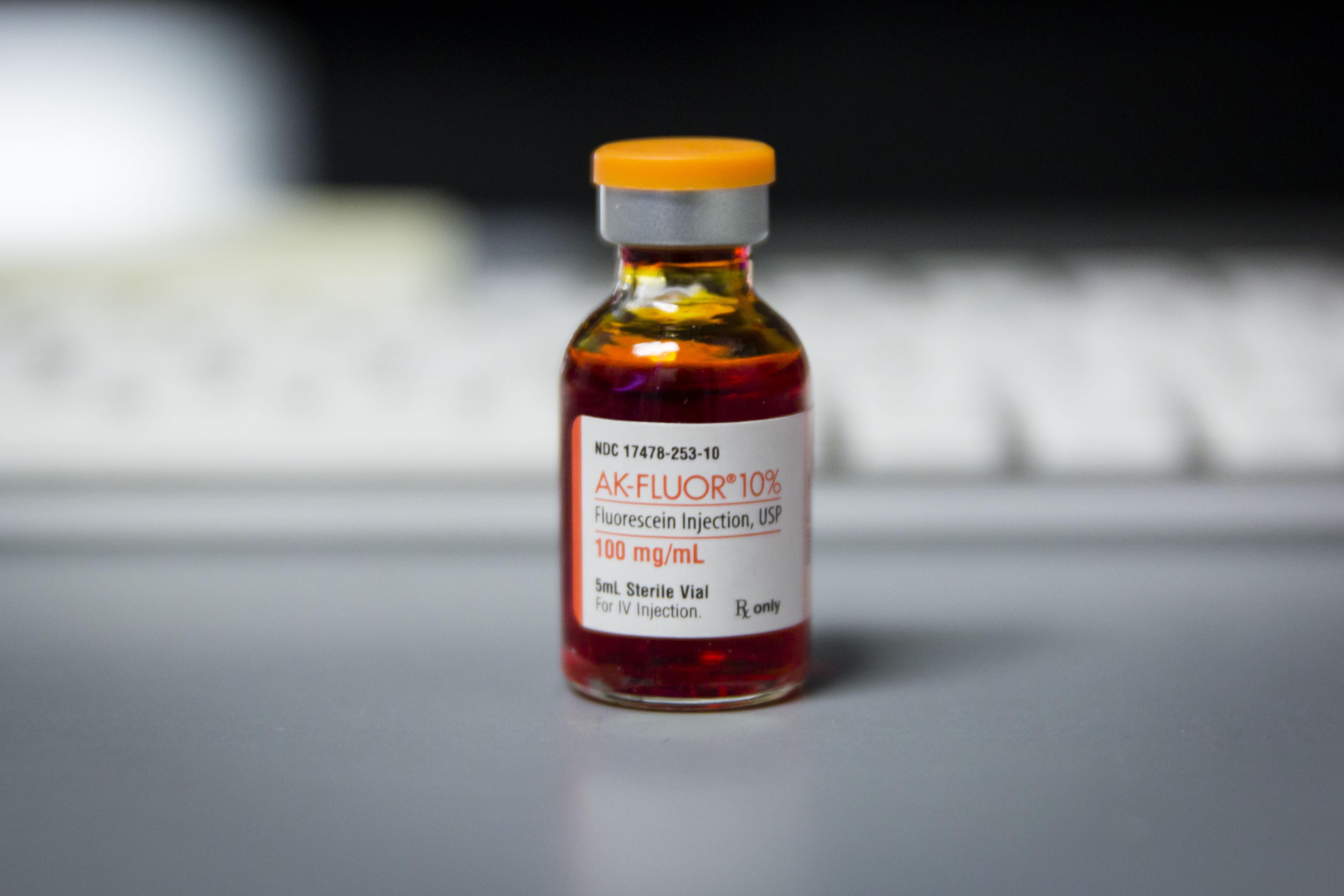
Fluorescein dye
Fluorescein angiography (FA) is useful for diagnosing retinal or optic nerve conditions, guiding laser surgery, and tracking the course of an existing condition. Fluorescein dye is injected into the arm, and photos are taken as it makes it way through the eye. Diabetic retinopathy, macular degeneration, retinal vein occlusions, and macular edema are some of the more common disorders diagnosed with FA.
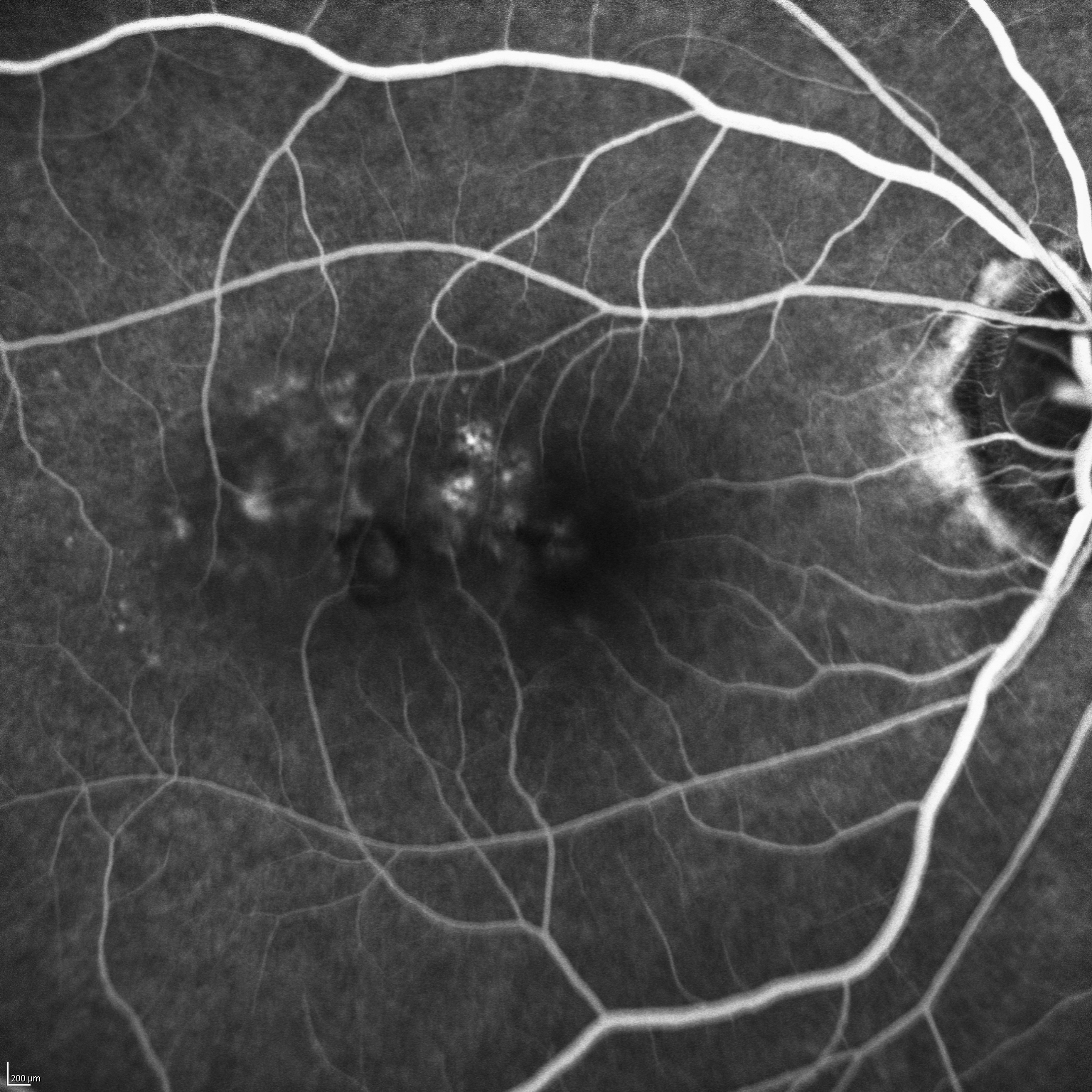
Early Phase FA – note the illumination of retinal blood vessels
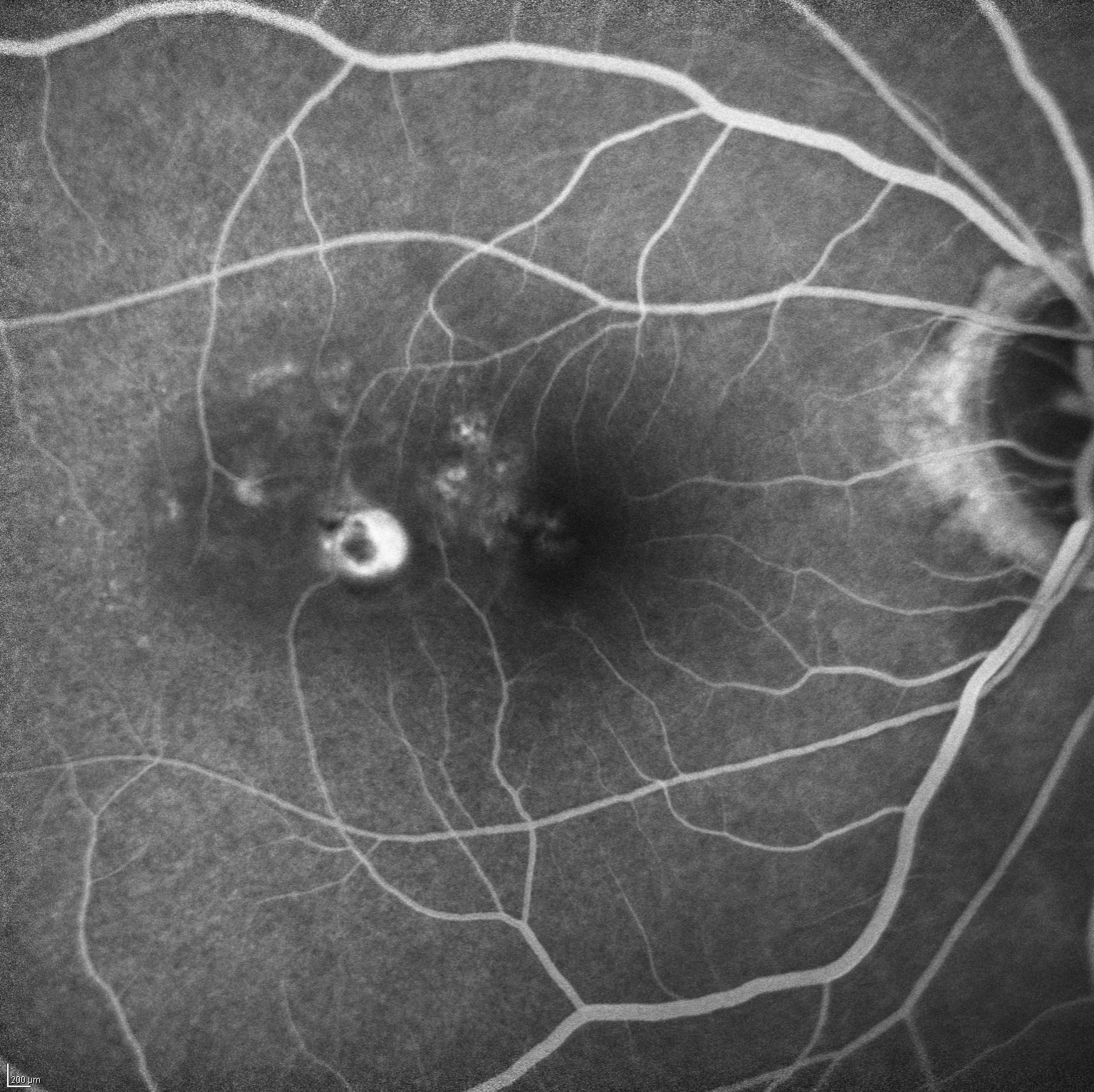
Late Phase FA – note the staining of the abnormal structures
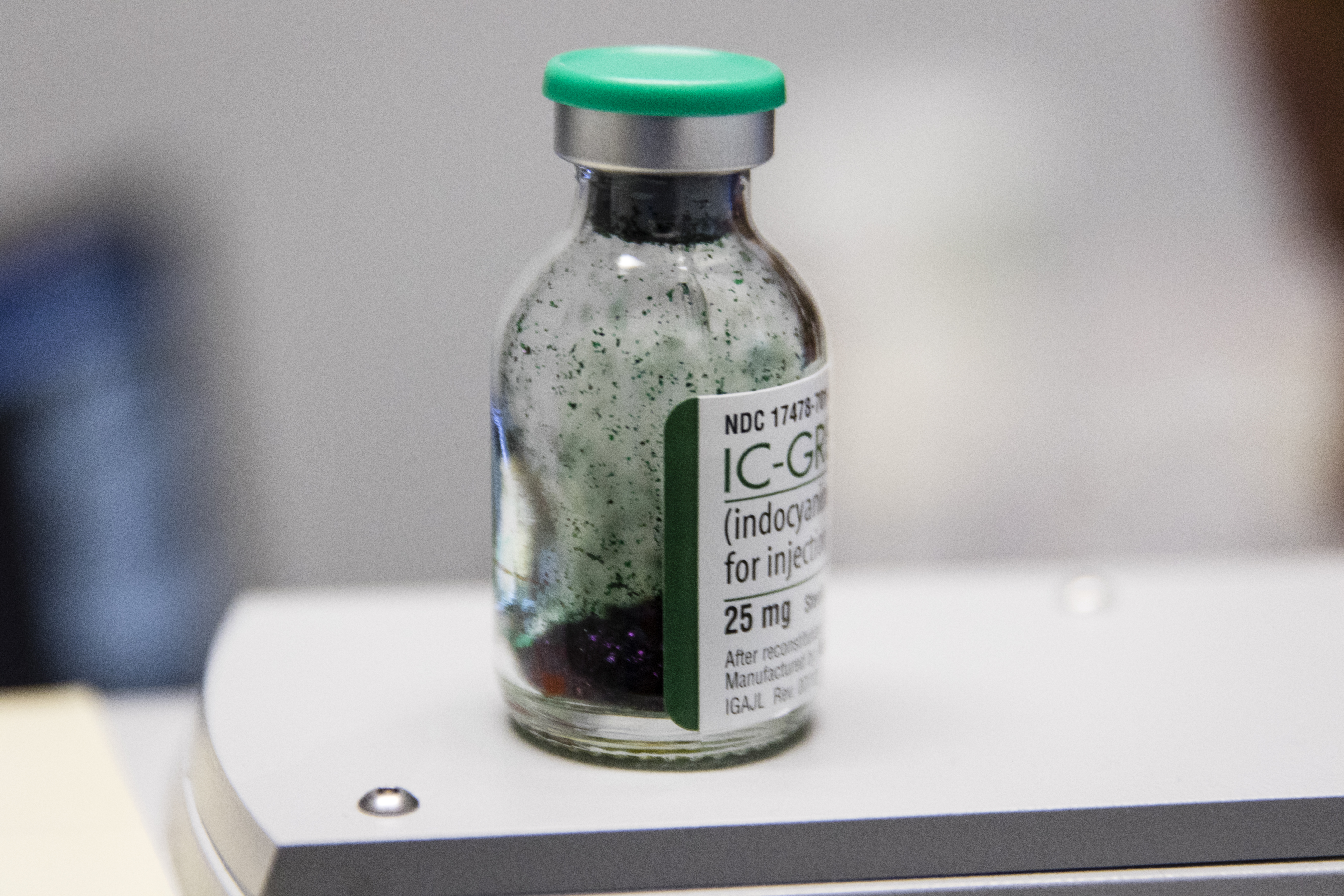
Indocyanine Green dye is usually in a powdered form and must be dissolved in sterile water before it is injected
Indocyanine green angiography
Indocyanine green angiography (ICGA) is used to view choroidal (the layer behind the retina) circulation. IGCA is excellent when patients have wet age-related macular degeneration. The abnormal blood vessels that grow underneath the retina can be blocked by blood or pigment with FA, but are visible with IGCA. ICGA is essential in the diagnosis of polypoidal choroidal vasculopathy (PCV).
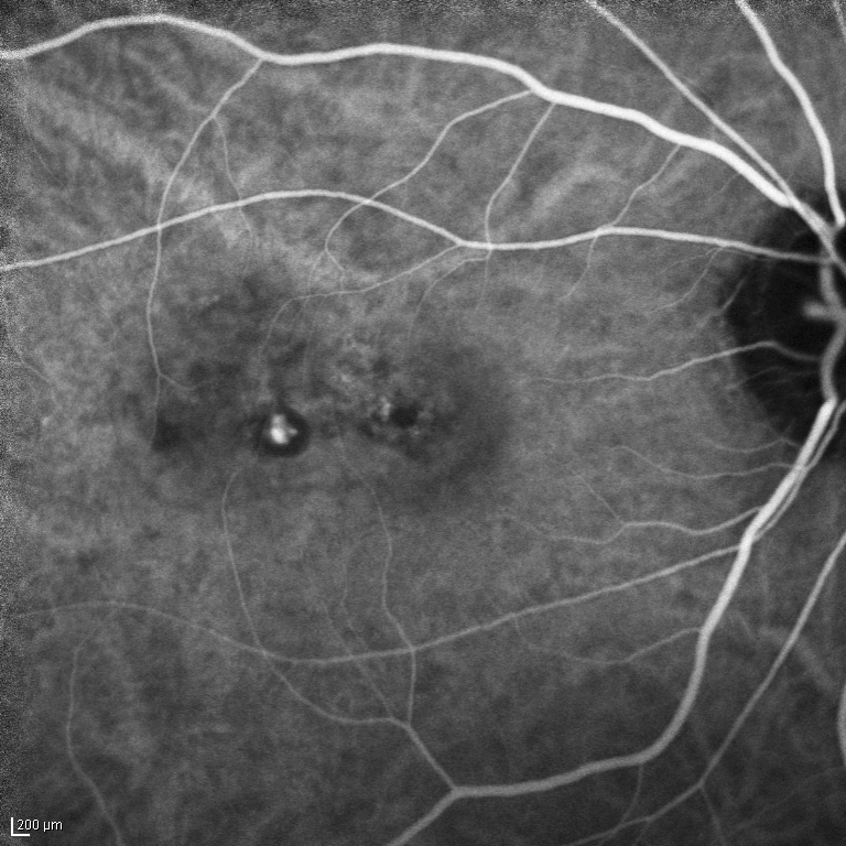
PCV diagnosed by ICGA – note illumination of blood vessels behind the retina
Optical coherence tomography
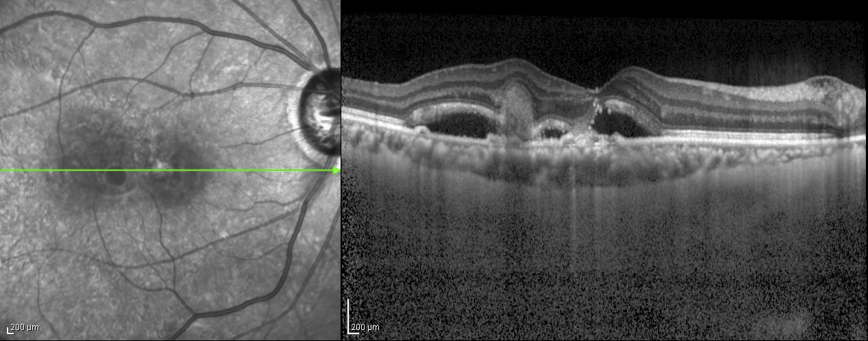
OCT produces cross-sectional images of the retina
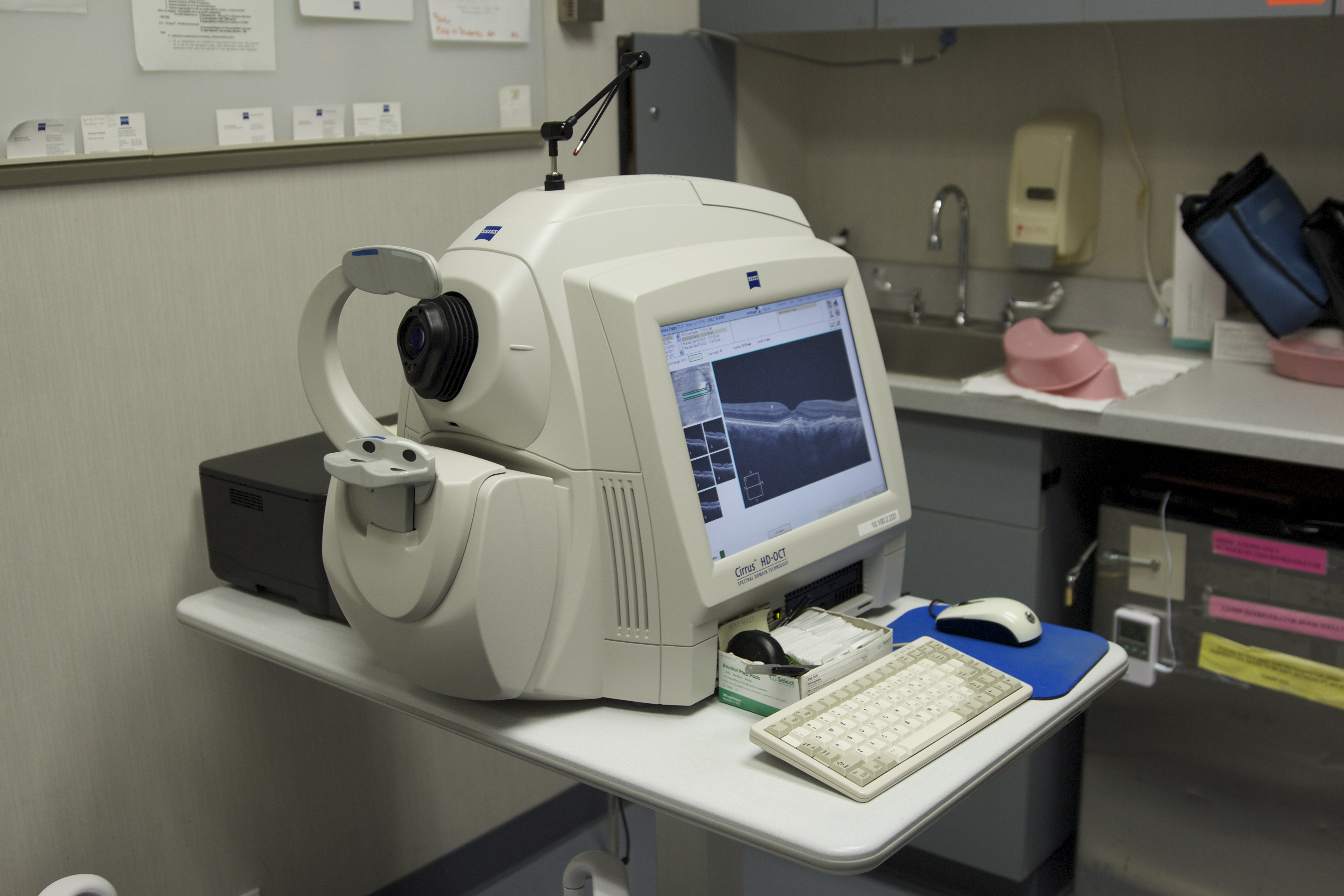
Cirrus HD-OCT (Zeiss)
Optical coherence tomography (OCT) is a noninvasive test where a scanning light beam provides a high-resolution cross section of the retina. It is useful in analyzing retinal conditions such as macular degeneration, macular edema, macular holes, macular puckers, diabetic retinopathy, and vein occlusions.
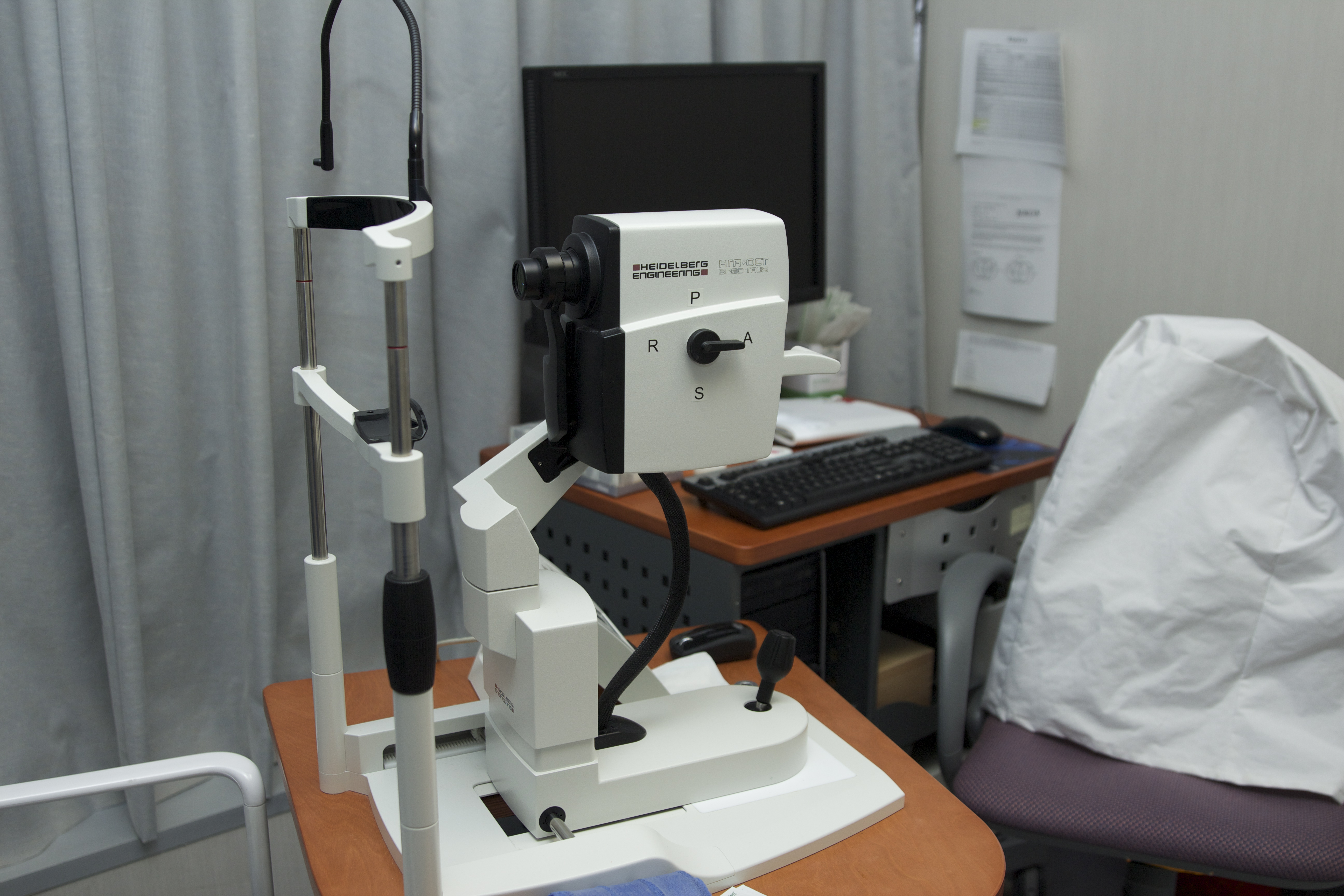
Spectralis HRA+OCT (Heidelberg Engineering, Inc)
Fundus Photography
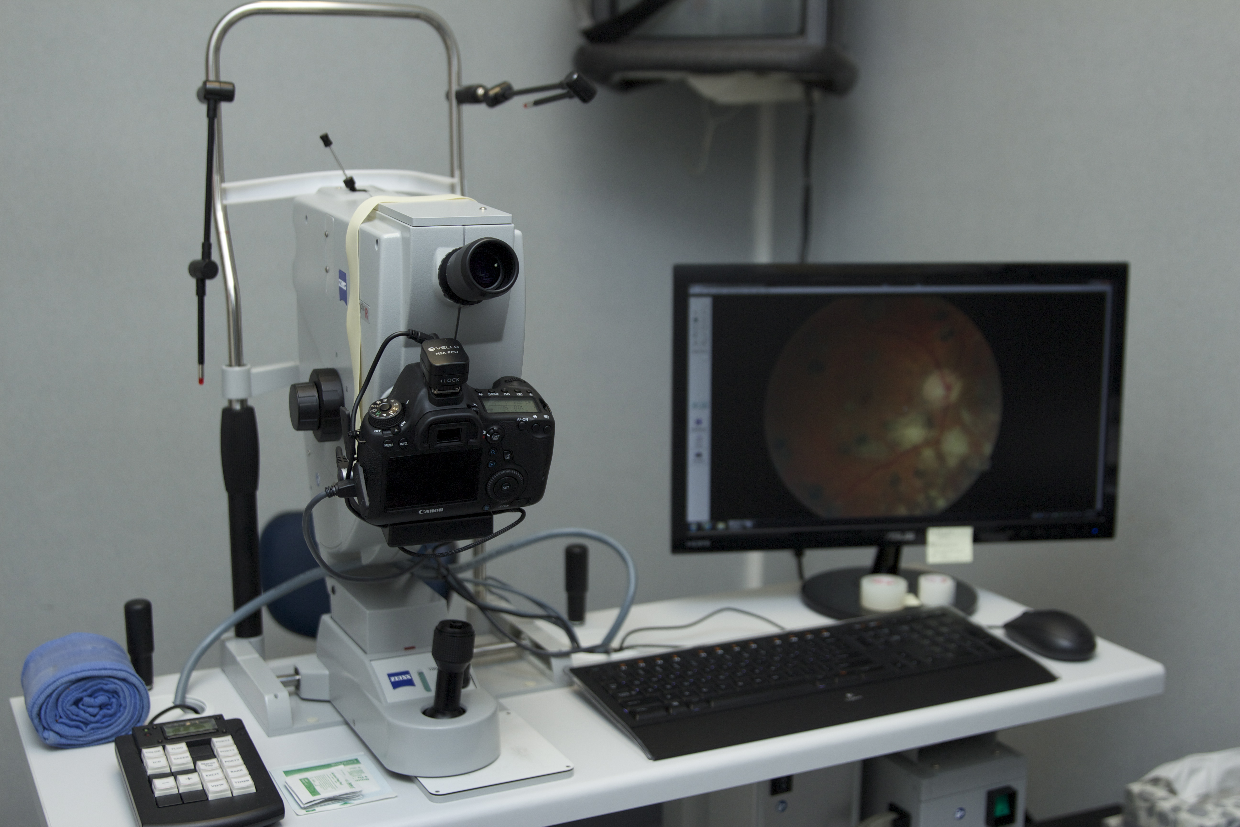
Fundus camera (left) with image on monitor (right)
A fundus photograph is a photographic image of the inside of the eye. The retina, optic disc, and retinal blood vessels can be viewed with a fundus photograph. Diseases such as glaucoma, retinal detachments, macular degeneration, and diabetic retinopathy can be monitored with fundus photographs.
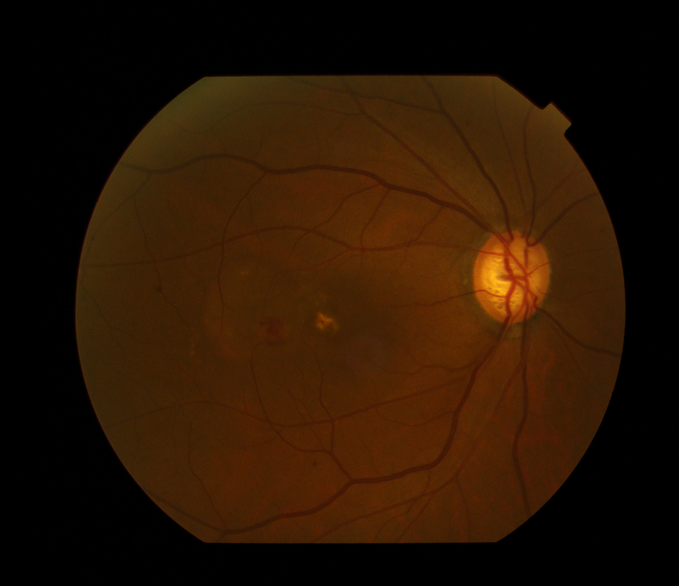
Fundus photograph of eye with PCV. Large polyp is visible in the center with hemorrhage
Ultrasound
Ultrasound uses sound waves to help visualize the back of the eye. These are the same waves used to check the fetus during pregnancy. Ultrasound provides a quick way of obtaining a clear view of the retina when either swelling, cataracts, or blood in the front of the eye is obscuring the physician’s view of the back of the eye.
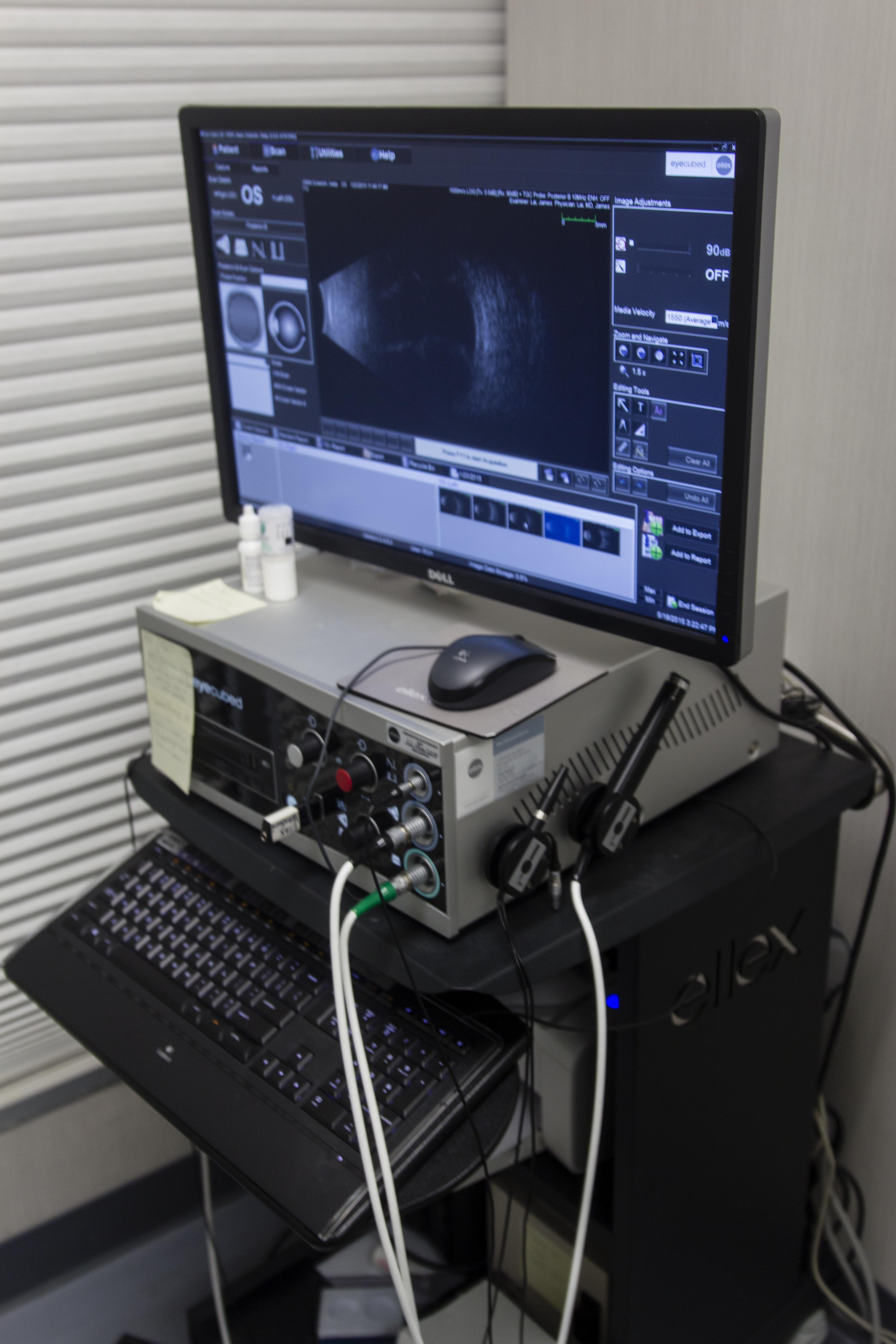
Eye Cubed Ultrasound (Ellex)
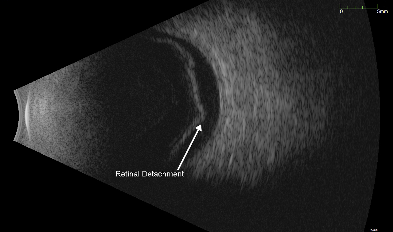
Retinal Detachment (arrow) imaged with ultrasound