Whether you are suffering from age-related macular degeneration, retinotherapy, floaters, or another disease entirely, you are probably wondering how your ophthalmologist will diagnose your condition. One process is through a test called an angiography.
How Does Angiography Work?
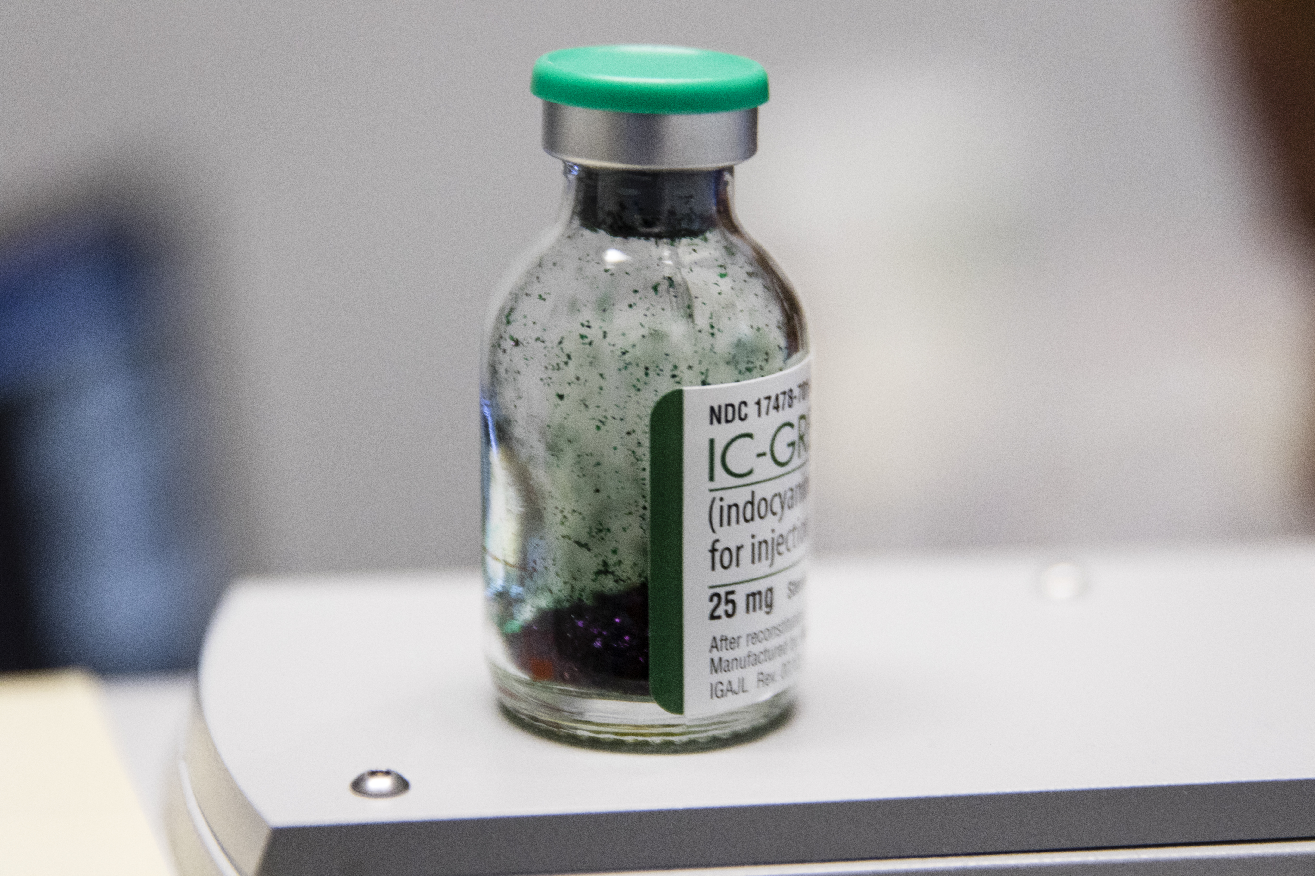
Indocyanine Green dye is a powder that must be diluted in sterile water before it is injected into your arm
During an angiography, a dye is injected into your body. This will be one of two dyes, either fluorescein or indocyanine green. Both dyes are safe and removed from the body by either the kidney or liver. However, if you are receving fluorescein angiography, your skin and urine usually turn orange for the next day. There are no other side effects, however.
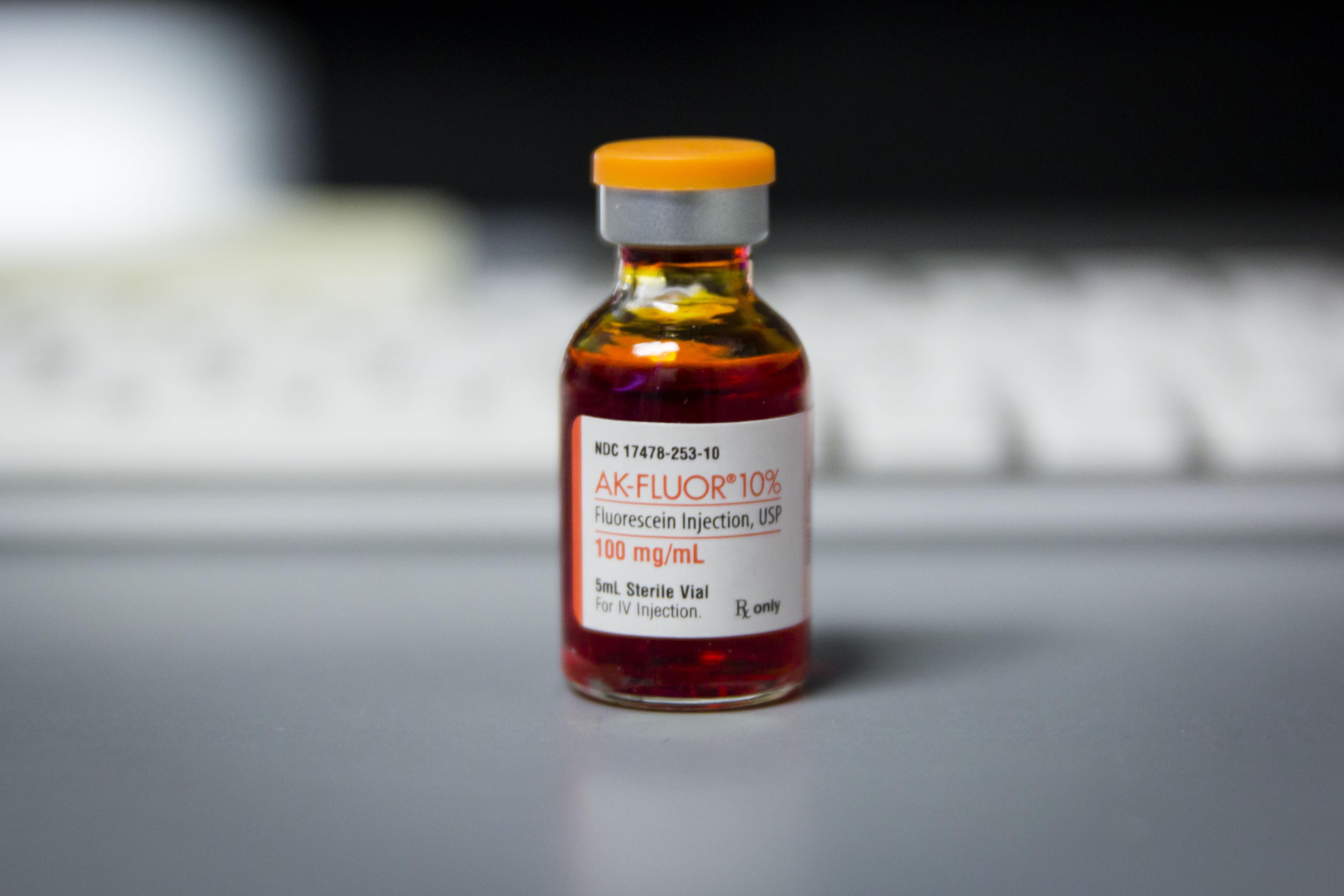
Fluorescein dye
Through a shot in your arm, the dye enters the your blood stream and travels throughout your body – including your eye. This is a painless process that lasts only a couple of minutes. As the dye passes through your retinal blood vessels, the photographer will take photographs that your doctor will analyze.
What Will an Angiography Tell Me?
Fluorescein angiography is the best way to examine retinal circulation. It can be used to diagnose almost any retinal condition, as well as keep doctors updated on the severity of your condition. It is therefore a very important test.
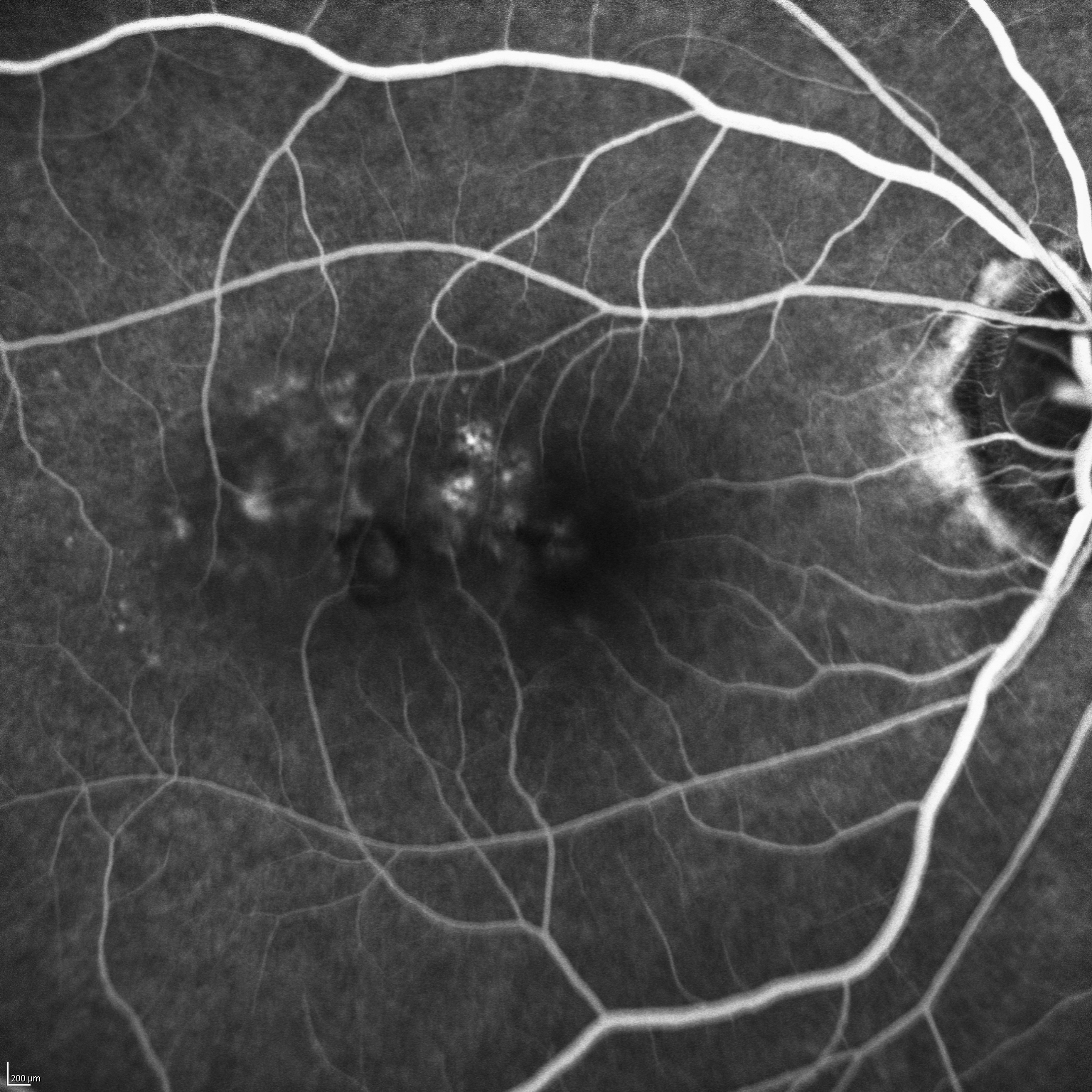
Early phase Fluorescein angiogram
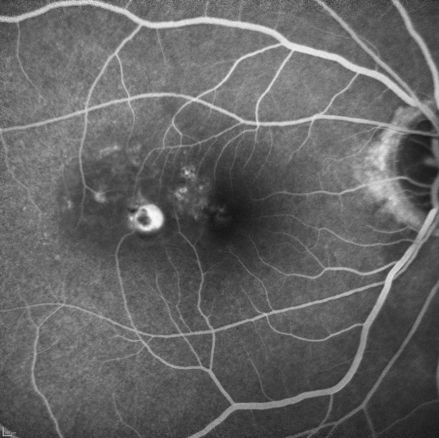
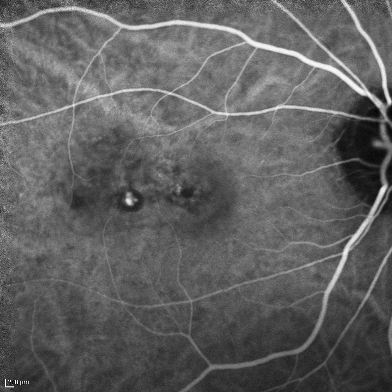
Late Phase Fluorescein Angiography
Indocyanine green angiography is most useful in diagnosing the wet form of AMD. The blood vessels that are underneath the retina and causing AMD may not be visible unless dyed with indocyanine green and photographed. In addition, this test is helpful when looking for tumors or inflammation.
Indocyanine Green Angiography
These tests are very important for you to maintain your eye health, and your doctor to deduce the correct diagnosis and provide the best treatment. To schedule an appointment, please call our office at (808) 487-8928.