Age-related macular degeneration (AMD) is a disease which affects the macula, the center of the retina that is responsible for your central vision. As the name implies, it develops in people as they get older. In fact, it is the leading cause of central vision loss in developed countries in people over the age of 55. If you think you might have macular degeneration and you are near the Honolulu, Hawaii area we can help.
Symptoms that patients may develop with AMD include:
- Blurred central vision
- Distortion of straight lines
- Blind spots in the central vision
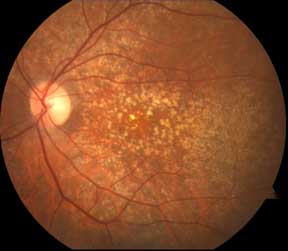
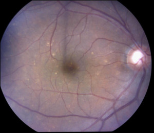
Drusen
Two Types of AMD
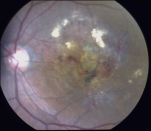
There are two different types of AMD, the more common dry form, and the less common wet form. In the dry form, which accounts for 90% of the AMD patients, drusen or yellowish aging deposits can develop under the retina. These deposits usually do not change vision and only a small percentage of people that have drusen will develop a more advanced form of macular degeneration that can be threatening to sharp, detailed vision. In the wet form, abnormal blood vessels (choroidal neovascularization) begin to grow under the retina. These abnormal blood vessels tend to bleed and leak under the retina, hence the name wet AMD. The bleeding and leakage lead to swelling and scarring in the macula with resultant loss of central vision. Although only 10% of patients with AMD have the wet form, they comprise over 90% of the patients with severe vision loss.
How is AMD diagnosed?
Your eye doctor can often diagnose AMD with a dilated eye examination. Often, additional testing such as fluorescein angiography, ICG angiography, and OCT will also be obtained to determine the type of macular degeneration that is present. These tests are also very useful in guiding treatment options.
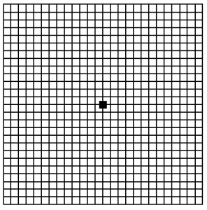
Once your doctor has diagnosed you with macular degeneration, it is helpful to use the following eye test to check your vision periodically in between doctor visits to see if you notice any changes in your vision. This eye test is called the Amsler Grid (see section on Amsler Grid). If you notice any changes such as wavy lines or areas that are gray/dark/discolored, it is important that you see your eye doctor as soon as possible.
Intravitreal injection Hawaii
What are the treatments?
While there are no cures for AMD, there are many new treatments that can help to control it.
For dry AMD, the Age-Related Eye Disease Study (AREDS) was an NIH-sponsored trial that demonstrated that certain high-dose vitamin supplementation could significantly reduce the progression of AMD. These vitamin formulations are available over the counter. We have a formation of these vitamins available Your physician will determine whether or not you would benefit from taking these vitamins. Furthermore, a diet high in green leafy vegetables, fish oils, and lutein, may also be beneficial. Smoking is a huge risk factor for the progression of both the dry and wet forms of AMD, so patients with macular degeneration must stop smoking.
For wet AMD, many treatment options are available. They include thermal laser treatment, photodynamic therapy with a non-thermal laser, and injections of medicines directly into the eye (intravitreal injections) to attack the choroidal neovascularization.
It is important to remember that the earlier the diagnosis, the more likely the treatment is to be successful. Check your vision in each eye periodically, preferably with an Amsler grid card, and notify your doctor if there is any change, especially distortion or blurriness. There are many treatment options available in the fight to preserve vision in patients with wet AMD. Your physician will be able to discuss with you which option is best for you.
Instructions for AMSLER GRID TESTING:
- Wear your reading glasses and cover one eye.
- Look at the center dot and keep your eye focused on this dot at all times.
- While looking at the center dot, make any notations of irregularly shaped boxes, wavy lines, distortions, discolorations.
- Repeat while covering the other eye.
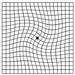
Amsler Grid Here is a Amlser Grid you can print out to use for self-monitoring. Below is an example of what an abnormal Amsler Grid may look like to someone that is taking this test that has retinal problems.
Abnormal Amsler Grid
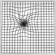
Abnormal Amsler Grid

