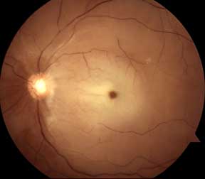
Arteries bring blood into the eyes while the veins take the blood out of the eyes back towards the heart. Sometimes a retinal artery can become occluded. In a way, one can think of a retinal artery occlusion as a “stroke or heart attack” of the eye. The patient will experience sudden painless loss of vision. If the blood flow is not restored within 90 minutes, the retina will begin to die and the vision loss will be permanent.
There are two main types of vein occlusions. A central artery occlusion occurs when the single main artery that supplies the retina becomes blocked. A branch artery occlusion occurs when a smaller branch off the main single main artery becomes blocked. Depending on how bad the occlusion is, both the central and branch artery occlusions can lead to severe ischemia and death of the retina.
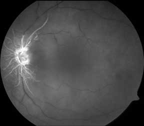
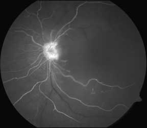
Your eye doctor will be able to diagnose this condition with a careful dilated examination. Additional testing with fluorescein angiograms and OCT are often obtained to assess the degree of damage to the retina.
There are no good treatments for retinal artery occlusions. Usually by the time that symptoms have developed, the nerve tissue in the retina has already died because it had not been receiving the necessary blood and nutrients. Patients with artery occlusions need to be monitored on a regular basis for the development of abnormal blood vessels that the eye sometimes develops in its attempt to resupply the damaged retina. If these abnormal blood vessels are found, your doctor will recommend a different laser treatment (panretinal photocoagulation) to get rid of these abnormal blood vessels
Retinal artery occlusions can be caused by many different reasons. Usually, the artery becomes blocked by an embolus. The most common sources of the emboli come from the heart or the carotids (arteries in one’s neck). Finding the underlying source of the embolus is critical to trying to prevent more emboli from forming and blocking more arteries to the eye, or even to the heart or brain. Your doctor will therefore often order additional tests to examine your heart (EKG, echocardiogram), your neck vessels (carotid Dopplers), and sometimes your blood to determine if you have any conditions that would predispose you for developing more emboli. Your primary care physician may also elect to place you on blood thinners to lower your risk of future occlusions.


Early Flourescein Angiogram Image of Central Retinal Artery Occlusion

Later Phase Fluorescein Angiogram of Central Retinal Artery Occlusion (CRAO)
