OCT uses a scanning light beam to acquire a high resolution cross section of the retina. This test is done in the office, and simply requires the patient to sit in front of a special camera. Since only light waves are used, no radiation is involved with this test. The information gathered by this test allows for accurate and reproducible images of the retina to be generated. It has been found to be particularly useful in the analysis of retinal conditions such as macular degeneration, macular edema, macular holes, macular puckers, diabetic retinopathy, and vein occlusions.
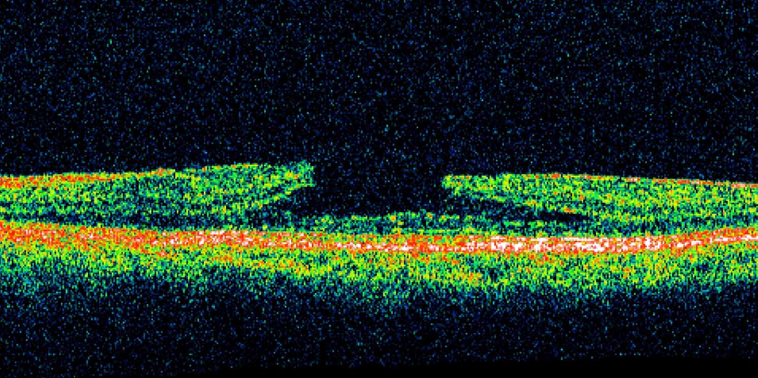
Abnormal OCT

Normal OCT