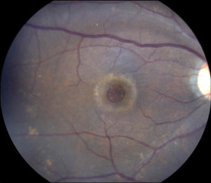
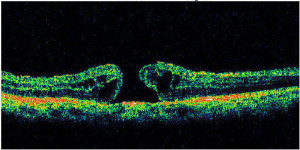
A macular hole is a small hole that develops in the center of the retina (macula). Because the macula is responsible for critical vision including reading, driving, and watching television, patients with a macular hole will notice that the center of their vision may be distorted or reduced while their peripheral vision is spared. Your doctor will often be able to diagnose the hole with a careful dilated examination. Often, additional testing with either a fluorescein angiogram or OCT may be obtained to better assess the size and chronicity of the hole.
Macular holes usually develop in otherwise healthy individuals as they get older. Holes will rarely close on their own. Typically when left untreated, the holes tend to get larger with resulting worsening central vision.
The only treatment available to close the hole is with vitrectomy surgery. A gas bubble is placed inside the eye to help push the hole closed. Because a gas bubble in the eye will always rise to the top (like bubbles rising to the surface when scuba diving), and because the macular hole is always directly in the back of the eye, patients are instructed to remain face down for several days to weeks after the operation. By remaining face down, it positions the bubble directly on the macular hole to help it close. The bubble gradually is replaced by the patient’s own tears, and the vision will slowly return. Rarely, silicone oil is used instead of a gas bubble.
The success rate for hole closure is over 90%. Most patients will experience an improvement in vision after the macular hole has closed. The distortion and blurred vision will likely improve, although it will never disappear altogether.
RCH participated in a national surgery study which helped study the surgical technique that is used today to treat this retinal condition. Macular holes have seen for over 100 years, and it has gone from a once untreatable condition to a condition that is treatable with surgery!
Macular Hole
OCT Scan of Macular Hole


